kw: book reviews, nonfiction, light, lighting, technology, infrastructure
When I began to read Brilliant: The Evolution of Artificial Light by Jane Brox, I was first prompted to consider the various technologies that have been used to light our homes and environment, from campfires to oil lamps to kerosene/mantle lamps to the electric filament bulb to fluorescents and LEDs. And what is next? Thinking about these things, and the continuing quest to produce more light and less heat, was behind this post of a couple days ago.
Ms Brox's book does indeed outline the progress of technologies used to create light, but I realized there is much more involved. There are three basic stages of light production, and each has been supported by a particular infrastructure, without which the light-producing device itself is of comparatively little use.
First, consider the technology of oil lamps, which are basically bowls with a wick and some melted or meltable oily substance. During the thousands (millions?) of years that this technology prevailed, there were the parallel technologies of oil/fat production and rendering, of producing the wicks, of passing the fire from place to place, and of starting the fire if yours has gone out and no near neighbor has any. This last was usually accomplished using a twirled hardwood rod, a dimple or hole in a piece of pine or other softwood, and some tinder.
When more volatile fuels such as light whale oil, kerosene and later coal gas came into use, primarily in Europe and America, there was a parallel development of chemical knowledge. Thus, while a technology for distributing fuels grew until it culminated in the neighborhood gas house and the piping systems that brought gaslight to homes and street corners, strikable matches and flint/steel strikers were also developed to make it easier to light one's lamps.
Finally, although the first incandescent electric light was energized in 1802 by Humphrey Davy, it took another 77 years of experimentation by many, including Edison's technicians, for a practical lamp to be produced. We give Edison credit for the bulb, but forget that his greater accomplishment was the infrastructure that made the bulb practical. The batteries of the day could only energize one of his bulbs for a short time, perhaps a few hours. Keeping that first bulb lit for fourteen hours until its glass envelope cracked was no mean feat, requiring the swapping of batteries in relays.
Thomas Edison envisioned a system of dynamos and underground, shielded wiring that would take care of the needs of a neighborhood at a time. His preference (almost a mania) for DC rather than AC current eventually led to his system being eclipsed by an AC infrastructure developed by George Westinghouse, based on Edison's but with the addition of transforming equipment so that power sent over a longer distance could be boosted to a higher voltage, reducing resistive losses in the wiring.
One characteristic of each successive infrastructure system has been increasing fragility. Thus, a few late chapters in the book cover the great blackout of 1965 and subsequent blackout events, and the efforts that are still being made to make "the grid" ever more robust and reliable. To meet the needs of the later 21st Century in America and Europe, at the very least, new trunk lines and newer switching equipment needs to be set up. It won't be cheap, but not doing it will eventually be hugely more costly. China, India and other major developing countries in Asia and Africa are watching our progress, so they can get their infrastructure right the first time.
With many business places lit entirely by fluorescent light, lighting consumes less than 8% of our energy budget. With many middle class homes using more and more CFL lamps, and a switch to LED's just beginning, this amount will decrease even more. But our use of light is the most visible manifestation of our electrical society. As we learn to make more light while using less energy to produce it, we also need to improve the efficiency of all our uses of energy.
I am reminded of something I learned forty years ago. AC power has this characteristic, that its use is less efficient if the voltage and current get out of phase. "Inductive" loads, such as electric motors, cause current to lag the voltage. "Capacitive" loads, and there are no simple examples, cause the current to lead instead. There are nearly no large electricity consuming devices that are capacitive, meaning that most industry operates with lagging current and its inherent inefficiencies. As it turns out, a type of electric motor called a synchronous motor acts as a large capacitive load. I don't understand the physics of it, so I can't explain why. Anyway, the primary market for large synchronous motors is major industrial plants that install them and keep them running just to balance the phase of their electricity, which significantly reduces their electric bill! I wonder if this would work at the house level. If I look at the transformers on the power poles up the street, I can see that some are accompanied by large capacitors, almost the size of the transformers, that the power company has installed to keep their own system in balance. Maybe there is a way to add a large capacitor or a synchronous motor in parallel with my furnace fan (the largest motor in the house). Don't hold your breath, but I do plan to look into this. Haven't given the matter any thought since 1970 until today!
The last chapter of the book is devoted to the loss of dark skies as we have gone to more and more street and exterior lighting. Not only is it inefficient, there is growing concern that it doesn't do as much as we thought to reduce crime. Perhaps just a little light is better than too much, while still being better than none at all. On my last visit to Japan, while my father-in-law was still living, I was told that he had installed most of the neighborhood's walkway lights. They were a series of single-tube fluorescent fixtures, and while they were nowhere near as bright as the lighting in my neighborhood, they were quite adequate for finding one's way around after dark. It was also easier to see the stars in the sky, which I much enjoy. His lights are probably very close to the amount of light we genuinely need, and a lot more economical than the way most American cities are lighted at present. Thanks to the author of Brilliant for such a thought-provoking book!
Showing posts with label light. Show all posts
Showing posts with label light. Show all posts
Monday, July 11, 2011


Friday, July 08, 2011
Light and heat in the front room
kw: analysis, light, technology
The way to make light, that is, to create photons, is to accelerate electrons. The oldest technology for doing this is to burn something. The hot carbon atoms in a candle flame emit light because the molecular collisions due to the heat knock electrons about. Also, carbon particles in the same flame emit light because the heat energy in the particles is carried by electron interactions, some of which produces "incandescent" photons. Neither process is particularly efficient, which is why lighting a room with candles, kerosene lamps, or gaslight creates a lot of heat. All the technologies of making light are driven by the need to do so at lower cost in power consumed, that is, in heat emitted.
I got to wondering how little heat I can produce while producing the light I want in my front room, by means of various technologies. There are two scenarios: (1) I am reading by myself; (2) We have guests. In the first case, I do quite nicely with one 18-watt CFL in a lamp that used to have a 75-watt tungsten bulb. The amount of light is equivalent, about 1100 lumens. The bulb's efficiency can be expressed as about 15 lumens per watt; the CFL's is about 62 lm/w.
In the second case, we add a stand light that has three 13-watt CFL's and a torchiere fitted with a screw fixture and a 23-watt CFL. That total of 80 watts of CFL's at 62 lm/w produces about 4,950 lumens. The same amount of light once needed 330 watts of incandescent bulbs.
But what if I had to use gaslight or candlelight? The former produces about 0.6 lm/w, the latter about half that. These watts are now counted as heat of burning. At 0.6 lm/w, 4,950 lumens requires more than 8,000 watts of heat. And candles? 16,000 watts seems an inconceivable amount! A heat pump type furnace consumes 3,000 watts to produce 10,000 watts of heat transfer. Even in winter, you'd drive everyone from the room in short order. This is why in the days of combustion-type lighting, the parlors were lit quite a bit more dimly than they are today.
And what of the future? Will LED's do better? The present state-of-the-art LED (so far only in the lab) produces 90 lm/w. I could light my room with 55 watts. That is a little better than today's 80 watts. LED's produce light by more cleverly accelerating electrons within certain semiconductor materials. The "white" ones in LED flashlights use a blue LED and a yellow wideband phosphor that converts part of the blue to green, yellow and red.
The theoretical maximum efficiency for an LED, using multi-colored emitters rather than fluorescent conversion, is either 200 or 250 lm/w, depending on the color mix, whether "flat white" or "center-hump". At 250 lm/w, I could produce 4,950 lumens using only 20 watts. Now, that's a goal worth shooting for.
The way to make light, that is, to create photons, is to accelerate electrons. The oldest technology for doing this is to burn something. The hot carbon atoms in a candle flame emit light because the molecular collisions due to the heat knock electrons about. Also, carbon particles in the same flame emit light because the heat energy in the particles is carried by electron interactions, some of which produces "incandescent" photons. Neither process is particularly efficient, which is why lighting a room with candles, kerosene lamps, or gaslight creates a lot of heat. All the technologies of making light are driven by the need to do so at lower cost in power consumed, that is, in heat emitted.
I got to wondering how little heat I can produce while producing the light I want in my front room, by means of various technologies. There are two scenarios: (1) I am reading by myself; (2) We have guests. In the first case, I do quite nicely with one 18-watt CFL in a lamp that used to have a 75-watt tungsten bulb. The amount of light is equivalent, about 1100 lumens. The bulb's efficiency can be expressed as about 15 lumens per watt; the CFL's is about 62 lm/w.
In the second case, we add a stand light that has three 13-watt CFL's and a torchiere fitted with a screw fixture and a 23-watt CFL. That total of 80 watts of CFL's at 62 lm/w produces about 4,950 lumens. The same amount of light once needed 330 watts of incandescent bulbs.
But what if I had to use gaslight or candlelight? The former produces about 0.6 lm/w, the latter about half that. These watts are now counted as heat of burning. At 0.6 lm/w, 4,950 lumens requires more than 8,000 watts of heat. And candles? 16,000 watts seems an inconceivable amount! A heat pump type furnace consumes 3,000 watts to produce 10,000 watts of heat transfer. Even in winter, you'd drive everyone from the room in short order. This is why in the days of combustion-type lighting, the parlors were lit quite a bit more dimly than they are today.
And what of the future? Will LED's do better? The present state-of-the-art LED (so far only in the lab) produces 90 lm/w. I could light my room with 55 watts. That is a little better than today's 80 watts. LED's produce light by more cleverly accelerating electrons within certain semiconductor materials. The "white" ones in LED flashlights use a blue LED and a yellow wideband phosphor that converts part of the blue to green, yellow and red.
The theoretical maximum efficiency for an LED, using multi-colored emitters rather than fluorescent conversion, is either 200 or 250 lm/w, depending on the color mix, whether "flat white" or "center-hump". At 250 lm/w, I could produce 4,950 lumens using only 20 watts. Now, that's a goal worth shooting for.
Monday, July 19, 2010
Make only photons you are going to use
kw: analysis, light, technology
I had a memory freeze yesterday while using a wind-up flashlight I have that uses LED's. I also have a hand-squeeze generator flashlight that uses a tiny incandescent bulb, but it has to be squeezed constantly to make any light. The LED one is would up for a half minute or a minute, then it works for about ten minutes.
I am glad for at least one kind of technological progress that makes better use of energy. I tracked down the luminous efficacy Wikipedia article while searching "lumens per watt", which contained the figures I needed to see historical progress, and speculate on the future.
First, a Lumen is a measure of the effective brightness of a light source. Its definition includes the spectral sensitivity of the human eye, which is at a maximum at a wavelength of 555nm, a yellowish-green. An ideal source, one that produced only 555nm photons, and made them with 100% efficiency, would have a luminous efficacy of 683 lumens per watt. By contrast, that now-obsolete 100 watt incandescent bulb that produces 1,400 lumens is producing 14 lumens per watt (lm/w), a total efficiency of just over 2%. Older office fluorescent tubes are 2.5 times as efficient, which is why the "standard" fluorescent tube has been the 40 watt size. Newer fluorescent tubes and compact fluorescent lamps (CFL's) are 5-6 times as efficient as the 100W bulb.
But I prefer a different standard of efficiency. The most efficient lamp available so far is the high-pressure sodium arc lamp, at 150 lm/w, or 22% total efficiency. But its strong yellow narrow-band spectrum produces a very ugly look to a scene, which is why they are only used for street lighting. The eye prefers broad-spectrum light, preferably full-spectrum white, that fills the 400-700nm visibility window. The most efficient possible "white", which contains only photons in the 400-700nm range, would yield 251 lumens per watt. That is 37% of the efficiency of a 555nm monochromatic source, but would be a lot easier on the eyes. Let us treat this source as the 100% standard in the discussion that follows.
By this standard, the old 100W incandescent bulb is 5.6% efficient. Although about 7% of the energy goes into visible photons, most of those are red, orange and some yellow, with very little being green and blue, so the overall efficiency is less than if it somehow produced an "equal energy white" spectrum. Incandescent technology got a small boost from the development of Halogen bulbs, which are filled not with vacuum (hmmm, kind of an oxymoron, that) but with argon and a little iodine. This allows the lamp to have a useful life while burning a little hotter, and the bulbs have 19 lm/w, an efficiency of 7.6%. That is almost 1.4 times as efficient as a bare 100W bulb. Now for some history.
The first light-producing technology was the campfire, soon followed by the candle. Both kinds of light source, converting BTU's to watts, produce about 0.3 lm/w, an efficiency near 0.1%. Two discoveries improved upon this, after the year 1800. First, the discovery of acetylene in the 1830s was followed within a generation by the development of gaslight for houses. It is three times as efficient as a candle or kerosene lamp. Then the gas mantle was developed in the 1880s, which is twice as efficient yet. Until Edison came along with the first carbon filament bulb (marginally more efficient), that was it. Tungsten began to be used for filaments in the 1920s, and the "early modern" incandescent bulb was on its way. Though fluorescent tubes were used in offices (similar time frame), they never became popular in homes because of their tendency to flicker. We just lived with lamps that wasted 94+% of their power.
CFL's of various shapes have about a 20-year history, in practical terms. It was about 1990 that they burgeoned into widespread use. They vary from 4-5 times as efficient as 100W incandescents. I have very few tungsten bulbs left in my house.
It is six years since I bought my first LED flashlight. It is a police-sized model with four D cells powering it, and 15 LED's. It is brighter than my old 2W flashlight, and the same four cells are still in it. The LED's are interesting. "White" LED's utilize a blue LED and a yellow phosphor that is efficiently excited by the blue wavelength. The yellow, consisting of moderately broadband red and green phosphors, mixes with the blue to produce a blue-white light. Newer screw-in LED bulbs in sizes from 4W to 8W, at prices of $70 or so, use the same technology.
There is a built-in inefficiency here, which I hope someone addresses. The conversion of blue to red and green entails a loss of about half the energy in the original blue light. I'd like to see a LED source that contained five or six LED's (or a multiple thereof), producing five or six wavelengths spread through the 400-700nm range. In other words, don't convert any photons, but produce only photons you are going to use. I suspect a nicely white lamp produced this way would have nearly twice the efficiency of the current LED's, perhaps 120-140 lm/w. This is in the range of 10x as efficient as a 100W bulb. How 'bout that? Light up a room using only 8-10 watts!
Then there is a further refinement. Just as most of the light from the 100W bulb is red and orange, the light from an LED source could be adjusted, but with a peak in the yellow-green instead of in the red. Such a "modulated" source might approach or exceed 150 lm/w, an efficiency of 60% or better. That is probably close to the ultimate that can be achieved, for a white-looking light. I can hardly wait.
I had a memory freeze yesterday while using a wind-up flashlight I have that uses LED's. I also have a hand-squeeze generator flashlight that uses a tiny incandescent bulb, but it has to be squeezed constantly to make any light. The LED one is would up for a half minute or a minute, then it works for about ten minutes.
I am glad for at least one kind of technological progress that makes better use of energy. I tracked down the luminous efficacy Wikipedia article while searching "lumens per watt", which contained the figures I needed to see historical progress, and speculate on the future.
First, a Lumen is a measure of the effective brightness of a light source. Its definition includes the spectral sensitivity of the human eye, which is at a maximum at a wavelength of 555nm, a yellowish-green. An ideal source, one that produced only 555nm photons, and made them with 100% efficiency, would have a luminous efficacy of 683 lumens per watt. By contrast, that now-obsolete 100 watt incandescent bulb that produces 1,400 lumens is producing 14 lumens per watt (lm/w), a total efficiency of just over 2%. Older office fluorescent tubes are 2.5 times as efficient, which is why the "standard" fluorescent tube has been the 40 watt size. Newer fluorescent tubes and compact fluorescent lamps (CFL's) are 5-6 times as efficient as the 100W bulb.
But I prefer a different standard of efficiency. The most efficient lamp available so far is the high-pressure sodium arc lamp, at 150 lm/w, or 22% total efficiency. But its strong yellow narrow-band spectrum produces a very ugly look to a scene, which is why they are only used for street lighting. The eye prefers broad-spectrum light, preferably full-spectrum white, that fills the 400-700nm visibility window. The most efficient possible "white", which contains only photons in the 400-700nm range, would yield 251 lumens per watt. That is 37% of the efficiency of a 555nm monochromatic source, but would be a lot easier on the eyes. Let us treat this source as the 100% standard in the discussion that follows.
By this standard, the old 100W incandescent bulb is 5.6% efficient. Although about 7% of the energy goes into visible photons, most of those are red, orange and some yellow, with very little being green and blue, so the overall efficiency is less than if it somehow produced an "equal energy white" spectrum. Incandescent technology got a small boost from the development of Halogen bulbs, which are filled not with vacuum (hmmm, kind of an oxymoron, that) but with argon and a little iodine. This allows the lamp to have a useful life while burning a little hotter, and the bulbs have 19 lm/w, an efficiency of 7.6%. That is almost 1.4 times as efficient as a bare 100W bulb. Now for some history.
The first light-producing technology was the campfire, soon followed by the candle. Both kinds of light source, converting BTU's to watts, produce about 0.3 lm/w, an efficiency near 0.1%. Two discoveries improved upon this, after the year 1800. First, the discovery of acetylene in the 1830s was followed within a generation by the development of gaslight for houses. It is three times as efficient as a candle or kerosene lamp. Then the gas mantle was developed in the 1880s, which is twice as efficient yet. Until Edison came along with the first carbon filament bulb (marginally more efficient), that was it. Tungsten began to be used for filaments in the 1920s, and the "early modern" incandescent bulb was on its way. Though fluorescent tubes were used in offices (similar time frame), they never became popular in homes because of their tendency to flicker. We just lived with lamps that wasted 94+% of their power.
CFL's of various shapes have about a 20-year history, in practical terms. It was about 1990 that they burgeoned into widespread use. They vary from 4-5 times as efficient as 100W incandescents. I have very few tungsten bulbs left in my house.
It is six years since I bought my first LED flashlight. It is a police-sized model with four D cells powering it, and 15 LED's. It is brighter than my old 2W flashlight, and the same four cells are still in it. The LED's are interesting. "White" LED's utilize a blue LED and a yellow phosphor that is efficiently excited by the blue wavelength. The yellow, consisting of moderately broadband red and green phosphors, mixes with the blue to produce a blue-white light. Newer screw-in LED bulbs in sizes from 4W to 8W, at prices of $70 or so, use the same technology.
There is a built-in inefficiency here, which I hope someone addresses. The conversion of blue to red and green entails a loss of about half the energy in the original blue light. I'd like to see a LED source that contained five or six LED's (or a multiple thereof), producing five or six wavelengths spread through the 400-700nm range. In other words, don't convert any photons, but produce only photons you are going to use. I suspect a nicely white lamp produced this way would have nearly twice the efficiency of the current LED's, perhaps 120-140 lm/w. This is in the range of 10x as efficient as a 100W bulb. How 'bout that? Light up a room using only 8-10 watts!
Then there is a further refinement. Just as most of the light from the 100W bulb is red and orange, the light from an LED source could be adjusted, but with a peak in the yellow-green instead of in the red. Such a "modulated" source might approach or exceed 150 lm/w, an efficiency of 60% or better. That is probably close to the ultimate that can be achieved, for a white-looking light. I can hardly wait.
Wednesday, March 03, 2010
Switching in less than a jiffy
kw: observations, science, physics, light, quantum theory
I'm reading a book about the historical development of quantum mechanics and entanglement. A review will appear in a couple more days; science histories take a while to read. I found fascinating the passionate debates engaged in by Bohr, Einstein, Schrodinger, Born, Ehrenfest, Heisenberg and others about what is really going on with quanta such as photons or electrons. Until deBroglie showed that the electron had a wave nature, it was not even considered a quantum.
As an objectivist (but not of the Rand variety), I am most compelled by things that actually happen. At root, a quantum behaves according to the kind of observation made upon it. The wave nature of photons, for example, is responsible for their ability to diffract when passing near an edge, to produce interference patterns, and to be refracted at the interface between differing media. The particle nature of photons is responsible for their ability to be detected by a photocell, a grain of silver chloride in photographic film, or even the retina of the eye.
The photons of light that form an image in your eye are focused as they enter the eye through the cornea, diffracted more or less by passing through a pupil of variable size, and focused more by the lens in the eye. For all these interactions, their wave nature prevails. Then, the energy of each photon is deposited in a dye molecule in a rod or cone cell, where it causes an electron to change its energy level. The electron then releases this energy into a nerve cell, which is now in a form that the brain can detect. The interaction with the electron depends on the photon's particle nature.
At one spot, the photon is behaving as a wave; at another less than 20mm away, it is behaving as a particle. At the speed photons travel through the eye (about 3/4 of their speed in vacuum), the "wave" interaction happens about 90 trillionths of a second before the "particle" interaction.
But that is from our point of view. What about the photon's "experience"? According to the theory of relativity, since a photon always travels at the speed of light, it experiences no passage of time; its "clock" is always stopped. From the time it is emitted, through its travels that possibly include reflections and refractions, until it is absorbed and moves one or more electrons about, the photon cannot experience anything but a timeless instant…speaking with gross anthropomorphism, of course! No matter "where" the points of emission and absorption may be, however far they may be separated, emission and absorption plus everything between are a single event.
There are several mysteries here, and though Heisenberg, Schrodinger and others developed ways of describing them mathematically, mysteries they remain. Yet the vision of every sighted creature (plus many other phenomena) depend on them, particularly on the dual nature of the photons.
I'm reading a book about the historical development of quantum mechanics and entanglement. A review will appear in a couple more days; science histories take a while to read. I found fascinating the passionate debates engaged in by Bohr, Einstein, Schrodinger, Born, Ehrenfest, Heisenberg and others about what is really going on with quanta such as photons or electrons. Until deBroglie showed that the electron had a wave nature, it was not even considered a quantum.
As an objectivist (but not of the Rand variety), I am most compelled by things that actually happen. At root, a quantum behaves according to the kind of observation made upon it. The wave nature of photons, for example, is responsible for their ability to diffract when passing near an edge, to produce interference patterns, and to be refracted at the interface between differing media. The particle nature of photons is responsible for their ability to be detected by a photocell, a grain of silver chloride in photographic film, or even the retina of the eye.
The photons of light that form an image in your eye are focused as they enter the eye through the cornea, diffracted more or less by passing through a pupil of variable size, and focused more by the lens in the eye. For all these interactions, their wave nature prevails. Then, the energy of each photon is deposited in a dye molecule in a rod or cone cell, where it causes an electron to change its energy level. The electron then releases this energy into a nerve cell, which is now in a form that the brain can detect. The interaction with the electron depends on the photon's particle nature.
At one spot, the photon is behaving as a wave; at another less than 20mm away, it is behaving as a particle. At the speed photons travel through the eye (about 3/4 of their speed in vacuum), the "wave" interaction happens about 90 trillionths of a second before the "particle" interaction.
But that is from our point of view. What about the photon's "experience"? According to the theory of relativity, since a photon always travels at the speed of light, it experiences no passage of time; its "clock" is always stopped. From the time it is emitted, through its travels that possibly include reflections and refractions, until it is absorbed and moves one or more electrons about, the photon cannot experience anything but a timeless instant…speaking with gross anthropomorphism, of course! No matter "where" the points of emission and absorption may be, however far they may be separated, emission and absorption plus everything between are a single event.
There are several mysteries here, and though Heisenberg, Schrodinger and others developed ways of describing them mathematically, mysteries they remain. Yet the vision of every sighted creature (plus many other phenomena) depend on them, particularly on the dual nature of the photons.
Thursday, June 04, 2009
It takes big eyes to see fine detail, part 4
kw: musings, physics, light, visibility
This continues a discussion I began here. I think the point is established that if you want to see fine detail, the solution is not a smaller "eye" but a larger one. But I have an error to correct.
In the prior post, I stated the Rayleigh criterion for the resolution of a diffraction-limited optical system, which is based on the Airy formula
x = 1.22 λl/d
where l/d is the focal ratio, such as 4:1 for an f/4 system.
I neglected to mention that this formula is only valid if the light path between the object and the viewing lens is vacuum (or air, to a close approximation). In the case of high-powered optical microscopes, the space between the object and the objective lens is filled with an oil that has the same index of refraction as the glass of the slide and of the lens, typically about 1.5. The symbol for refractive index is n, so in an optical microscope with an oil-immersion objective, the formula is
x = 1.22 λl/nd
This replacement of λ with λ/n resolves the difference I'd stated between the Rayleigh criterion and the Abbe limit: 0.866/1.5 = 0.58, which means that an oil-immersed lens with N.A. = 1.4 (f/0.71) will resolve details as small as 0.58λ, which is 0.35µ for incandescent light (effective λ = 0.6µ) and 0.28µ for daylight (effective λ = 0.48µ).
My calculations for ultraviolet viewing remain unchanged because immersion oils absorb UV light.
Now, before we consider even smaller wavelengths, there is a technology that can resolve very small details for certain objects, using visible light. That is Near-field Scanning Optical Microscopy, or NSOM.
A very (very!) smooth sample can be scanned with a narrow glass fiber tip, just a few nanometers across. As long as the fiber tip is within a few nm of the surface being scanned, the spot of light will effectively be very much smaller than a wavelength. In practice, resolutions of around 10nm have been achieved. This is fifty times as good as imaging with lens optics. This technology is not for the faint of heart or poverty of pocketbook. Just drawing a glass fiber so it necks down to 3-5 nm diameter is a challenging task. Handling it to mount it and use it without breaking it…tricky. If you don't need the optical response, electron microscopes are cheaper, both SEM and TEM.
The concept to grasp for what follows is that of disturbance. We don't think of it this way, but even using visible light to view an object disturbs the object. The way light is reflected requires disturbing the outer electrons of a substance, which either absorb or re-emit the photons that bang into them. Visible photon energies are low, however, and don't typically cause atoms to be shifted out of position. Even x-rays and electrons a thousand times as energetic as visible photons seldom force atoms to jump aside.
But the electron bombardment, in particular, cause a disturbance in electron orbits. When a high-powered electron microscope is used to image atoms, we are seeing them slightly distorted, because what we are seeing is an image of the threshold position of the electron cloud around each atom's nucleus, at some equilibrium position as blown by the "wind" of electrons we are viewing with.
Is viewing the atoms, or their outer electron clouds, the ultimate achievable? Not at all. Hitting a small enough sample with a sufficiently dense electron beam, we can strip off the outer electrons, ionizing the atoms in the sample, but the ions will quickly neutralize by grabbing electrons from a cloud that builds up around the sample. Ion imaging may have its place, but I haven't heard of it being used. Nonetheless, there may be a way to use such a technique to probe various electron orbitals. X-rays are more typically used to do that, however, and the technology is well understood. We won't go further with that here.
Another reason for disturbing atoms enough to ionize them is related to probing the mechanics of the structure of a single atom's electron cloud. In this small realm, the order and spacing of each electron's orbital cloud are governed by quantum mechanics. For such studies to be meaningful, the atoms need to be isolated, so charged gases and plasmas are used. Atoms so disturbed emit and absorb electromagnetic radiation of all kinds, so this is the realm of spectroscopy, which covers a vast range of energies, from quite low (medium infrared starting at about 3µ) through visible and ultraviolet to x-rays with wavelengths of less than 1nm and energies of 100 KeV or more.
The next barrier is the atomic nucleus. What does it take to disturb it enough to learn about its innards? Ernest Rutherford began to show the way by bombarding gold foil with alpha particles, which are totally ionized helium nuclei. He was throwing one kind of nucleus at another, though he didn't know it. The α particles had an energy near 4 MeV, and when one came close to a gold nucleus, it bounced right back, or to the side. So four million volts isn't enough to do more than get a rough fix on the location of a nucleus.
As it turns out, once the proton was discovered (it is a bare hydrogen nucleus), it was found to weigh about 1,800 times as much as an electron. It takes many MeV to do more than make a proton bounce around like a smacked ping pong ball. Today's particle physics, developed since the 1950s, is in the business of finding out how the components of protons work together. It takes millions of MeV to have much chance of cracking open a proton (AKA p+), and its energetic components are very shy, splitting into showers of other particles within 10-24 second.
The diameter of a nucleus is so small that, it has been often written, its size compared to the electron cloud is similar to a grain of sand hovering at the center of a cathedral or airplane hanger. An iron atom is about 10-10m in diameter, and its nucleus is a bit smaller than 10-13m, a thousand times smaller.
How big a machine is required to "see" details this small, and smaller? As it turns out, the finer the details you want to see, the bigger the machine it takes. Particle Accelerators are the tool we need to produce a probe that can see inside a proton. Accelerators are of two types: linear and circular. An ordinary TV set, and a small electron microscope, are both accelerators of the linear type, that accelerate electrons to energies of 30-50 KeV. Early accelerators that fit in a lab with a high ceiling, known as Cockcroft-Walton accelerators, could produce electrons with energies of a few MeV, and they are 4-5m tall. Both these kinds of accelerator are one-push types: make lots of volts and let electrons "fall" from one end to the other.
Higher energies are produced by letting the accelerated electrons fly through a hole and giving them another push with radio waves. The biggest of these, the Stanford Linear Accelerator (SLAC) imparts energies up to 50 GeV to electrons. But linear is a one-shot kind of technology. Once the electrons fly out the end, you're done with them. Much higher energies are had by running particles in circles, so you can push them over and over again, then divert them to hit your target.
The modern tool for this is the synchrotron. The old Cosmotron was 23m in diameter and imparted a bit more than 3 GeV to protons. The main synchrotron that accelerates electrons, the LEP, gives them the same energy as SLAC, but it can run beams in opposite directions so as to crash them head-on, for more energetic collisions. It can do the same with positrons, or with positrons one way and electrons the other. Lotsa possibilities! But protons, being 1,800 times as heavy, achieve much higher energies. The largest proton accelerator, the LHC (which operated briefly late last year, and is now being repaired!), should achieve 7 TeV (million MeV) per proton. But the LHC is designed to accelerate heavy nuclei containing many protons and neutrons, for purposes of learning more about how the quarks and gluons that make up a whole nucleus interact. The LHC is 8,500m in diameter, or 8km.
Putting the few figures I've given above with others, I produced this chart:
 In the legend, p+ is for proton and e- is for electron. For electrons, getting to a certain energy takes about the same size machine, whether linear or circular. Circular machines have losses, however, because turning the electrons in a circle causes bursts of electromagnetic energy to leak away. This accounts for the somewhat different scale between the red and blue lines.
The green line shows proton energies in a few synchrotrons. The straightness of the line allows us to determine a formula, for gaining more energy if our investigations in the future require it:
Emax = 53.2 MeV x Diameter1.3, for Diameter in meters.
Though we are producing particles with TeV scale energies, more is always desired. The 10-24s time scale of quark-gluon interactions requires a wavelength of 3x10-16m, or a particle energy of 41 GeV. In practice, it takes many times this energy, most of which is used to create "resonance" particles that spall away, so some is left over to push a couple of quarks a few trillionths of a meter apart so you can get a reaction you can measure.
How much energy is "enough"? Some Cosmic rays are more energetic than the output of the biggest accelerator we could build on earth. Let us use the formula above to calculate the energy from an accelerator 14,700km in diameter: we get 1.1x10¹⁷ eV, or 110,000 TeV. The most powerful cosmic ray so far detected had an energy of 3x10¹⁸ eV, and an accelerator to produce such particles would need to be 4 million km in diameter.
Until new technologies allow more efficient circular accelerators, then, we are limited to some thousands of TeV. The wavelength of ~100,000 TeV particles is as short as 10-22m, and they can interact on time scales as brief as 10-30 second. And that's the finest resolution achievable from machines that fit onto the surface of the earth.
This continues a discussion I began here. I think the point is established that if you want to see fine detail, the solution is not a smaller "eye" but a larger one. But I have an error to correct.
In the prior post, I stated the Rayleigh criterion for the resolution of a diffraction-limited optical system, which is based on the Airy formula
x = 1.22 λl/d
where l/d is the focal ratio, such as 4:1 for an f/4 system.
I neglected to mention that this formula is only valid if the light path between the object and the viewing lens is vacuum (or air, to a close approximation). In the case of high-powered optical microscopes, the space between the object and the objective lens is filled with an oil that has the same index of refraction as the glass of the slide and of the lens, typically about 1.5. The symbol for refractive index is n, so in an optical microscope with an oil-immersion objective, the formula is
x = 1.22 λl/nd
This replacement of λ with λ/n resolves the difference I'd stated between the Rayleigh criterion and the Abbe limit: 0.866/1.5 = 0.58, which means that an oil-immersed lens with N.A. = 1.4 (f/0.71) will resolve details as small as 0.58λ, which is 0.35µ for incandescent light (effective λ = 0.6µ) and 0.28µ for daylight (effective λ = 0.48µ).
My calculations for ultraviolet viewing remain unchanged because immersion oils absorb UV light.
Now, before we consider even smaller wavelengths, there is a technology that can resolve very small details for certain objects, using visible light. That is Near-field Scanning Optical Microscopy, or NSOM.
A very (very!) smooth sample can be scanned with a narrow glass fiber tip, just a few nanometers across. As long as the fiber tip is within a few nm of the surface being scanned, the spot of light will effectively be very much smaller than a wavelength. In practice, resolutions of around 10nm have been achieved. This is fifty times as good as imaging with lens optics. This technology is not for the faint of heart or poverty of pocketbook. Just drawing a glass fiber so it necks down to 3-5 nm diameter is a challenging task. Handling it to mount it and use it without breaking it…tricky. If you don't need the optical response, electron microscopes are cheaper, both SEM and TEM.
The concept to grasp for what follows is that of disturbance. We don't think of it this way, but even using visible light to view an object disturbs the object. The way light is reflected requires disturbing the outer electrons of a substance, which either absorb or re-emit the photons that bang into them. Visible photon energies are low, however, and don't typically cause atoms to be shifted out of position. Even x-rays and electrons a thousand times as energetic as visible photons seldom force atoms to jump aside.
But the electron bombardment, in particular, cause a disturbance in electron orbits. When a high-powered electron microscope is used to image atoms, we are seeing them slightly distorted, because what we are seeing is an image of the threshold position of the electron cloud around each atom's nucleus, at some equilibrium position as blown by the "wind" of electrons we are viewing with.
Is viewing the atoms, or their outer electron clouds, the ultimate achievable? Not at all. Hitting a small enough sample with a sufficiently dense electron beam, we can strip off the outer electrons, ionizing the atoms in the sample, but the ions will quickly neutralize by grabbing electrons from a cloud that builds up around the sample. Ion imaging may have its place, but I haven't heard of it being used. Nonetheless, there may be a way to use such a technique to probe various electron orbitals. X-rays are more typically used to do that, however, and the technology is well understood. We won't go further with that here.
Another reason for disturbing atoms enough to ionize them is related to probing the mechanics of the structure of a single atom's electron cloud. In this small realm, the order and spacing of each electron's orbital cloud are governed by quantum mechanics. For such studies to be meaningful, the atoms need to be isolated, so charged gases and plasmas are used. Atoms so disturbed emit and absorb electromagnetic radiation of all kinds, so this is the realm of spectroscopy, which covers a vast range of energies, from quite low (medium infrared starting at about 3µ) through visible and ultraviolet to x-rays with wavelengths of less than 1nm and energies of 100 KeV or more.
The next barrier is the atomic nucleus. What does it take to disturb it enough to learn about its innards? Ernest Rutherford began to show the way by bombarding gold foil with alpha particles, which are totally ionized helium nuclei. He was throwing one kind of nucleus at another, though he didn't know it. The α particles had an energy near 4 MeV, and when one came close to a gold nucleus, it bounced right back, or to the side. So four million volts isn't enough to do more than get a rough fix on the location of a nucleus.
As it turns out, once the proton was discovered (it is a bare hydrogen nucleus), it was found to weigh about 1,800 times as much as an electron. It takes many MeV to do more than make a proton bounce around like a smacked ping pong ball. Today's particle physics, developed since the 1950s, is in the business of finding out how the components of protons work together. It takes millions of MeV to have much chance of cracking open a proton (AKA p+), and its energetic components are very shy, splitting into showers of other particles within 10-24 second.
The diameter of a nucleus is so small that, it has been often written, its size compared to the electron cloud is similar to a grain of sand hovering at the center of a cathedral or airplane hanger. An iron atom is about 10-10m in diameter, and its nucleus is a bit smaller than 10-13m, a thousand times smaller.
How big a machine is required to "see" details this small, and smaller? As it turns out, the finer the details you want to see, the bigger the machine it takes. Particle Accelerators are the tool we need to produce a probe that can see inside a proton. Accelerators are of two types: linear and circular. An ordinary TV set, and a small electron microscope, are both accelerators of the linear type, that accelerate electrons to energies of 30-50 KeV. Early accelerators that fit in a lab with a high ceiling, known as Cockcroft-Walton accelerators, could produce electrons with energies of a few MeV, and they are 4-5m tall. Both these kinds of accelerator are one-push types: make lots of volts and let electrons "fall" from one end to the other.
Higher energies are produced by letting the accelerated electrons fly through a hole and giving them another push with radio waves. The biggest of these, the Stanford Linear Accelerator (SLAC) imparts energies up to 50 GeV to electrons. But linear is a one-shot kind of technology. Once the electrons fly out the end, you're done with them. Much higher energies are had by running particles in circles, so you can push them over and over again, then divert them to hit your target.
The modern tool for this is the synchrotron. The old Cosmotron was 23m in diameter and imparted a bit more than 3 GeV to protons. The main synchrotron that accelerates electrons, the LEP, gives them the same energy as SLAC, but it can run beams in opposite directions so as to crash them head-on, for more energetic collisions. It can do the same with positrons, or with positrons one way and electrons the other. Lotsa possibilities! But protons, being 1,800 times as heavy, achieve much higher energies. The largest proton accelerator, the LHC (which operated briefly late last year, and is now being repaired!), should achieve 7 TeV (million MeV) per proton. But the LHC is designed to accelerate heavy nuclei containing many protons and neutrons, for purposes of learning more about how the quarks and gluons that make up a whole nucleus interact. The LHC is 8,500m in diameter, or 8km.
Putting the few figures I've given above with others, I produced this chart:
 In the legend, p+ is for proton and e- is for electron. For electrons, getting to a certain energy takes about the same size machine, whether linear or circular. Circular machines have losses, however, because turning the electrons in a circle causes bursts of electromagnetic energy to leak away. This accounts for the somewhat different scale between the red and blue lines.
The green line shows proton energies in a few synchrotrons. The straightness of the line allows us to determine a formula, for gaining more energy if our investigations in the future require it:
Emax = 53.2 MeV x Diameter1.3, for Diameter in meters.
Though we are producing particles with TeV scale energies, more is always desired. The 10-24s time scale of quark-gluon interactions requires a wavelength of 3x10-16m, or a particle energy of 41 GeV. In practice, it takes many times this energy, most of which is used to create "resonance" particles that spall away, so some is left over to push a couple of quarks a few trillionths of a meter apart so you can get a reaction you can measure.
How much energy is "enough"? Some Cosmic rays are more energetic than the output of the biggest accelerator we could build on earth. Let us use the formula above to calculate the energy from an accelerator 14,700km in diameter: we get 1.1x10¹⁷ eV, or 110,000 TeV. The most powerful cosmic ray so far detected had an energy of 3x10¹⁸ eV, and an accelerator to produce such particles would need to be 4 million km in diameter.
Until new technologies allow more efficient circular accelerators, then, we are limited to some thousands of TeV. The wavelength of ~100,000 TeV particles is as short as 10-22m, and they can interact on time scales as brief as 10-30 second. And that's the finest resolution achievable from machines that fit onto the surface of the earth.
Wednesday, June 03, 2009
It takes big eyes to see fine detail, part 3
kw: musings, physics, light, visibility
This is the third in a series of posts that began here. In that post, it was first determined that the resolution in the sensor plane depends only on the focal ratio (f-stop or f/number). A f/4 lens-sensor (film, retina, or digital camera chip) system will record ("see") details no finer than 3µ apart when the effective wavelength is 0.6µ, no matter how large or small the lens is. Thus a physically larger camera with f/4 optics will record more total detail than a smaller one. All this was based on "ordinary" photography and viewing, with the camera-to-subject distance being greater than the lens-to-sensor distance.
In the second post, this situation was reversed, and we examined photomicrography and the limits of optical magnification. The absolute minimum f/number for any lens is f/0.5, and the practical limit is f/0.6, while the standard for microscope optics (other than rare special products) is f/0.71. At that ratio, the lens diameter is 1.4 times the distance between the object being viewed and the optical center of the lens. The smallest details such a system can view are 0.52µ apart using incandescent light (effective wavelength 0.6µ) and 0.42µ apart when using a bluer, more daylight-like light (0.48µ).
Microscopists quote the Abbe Limit, which claims resolution close to half the wavelength for a f/0.71 system, but I have not been convinced by my own experience. The Rayleigh criterion I rely on predicts instead 0.866 times the effective wavelength for an f/0.71 system. Whichever criterion we prefer, though, how do we see stuff smaller than that? We use "light" with a shorter wavelength.
Two technologies are currently used to image microscopically using wavelengths shorter than 0.4µ or 400nm (From this point, wavelengths will be expressed in nanometers, since a micron (µ) is large on these scales).
I was familiar with optical aberrations because of my interest in astronomy and telescopes; a friend of my father's was making a refracting telescope, and explained why it takes careful matching of the curvatures of four surfaces, on two pieces of glass, to get a lens that has acceptable correction of both chromatic and spherical aberration. A single spherical surface does not focus parallel light to a single point, but to a focal ray that is elongated along the optical axis. For a single wavelength of light, two spherical surfaces can correct most of this spherical aberration. Multi-wavelength (and thus multi-colored) light is also focused at different distances, and it takes a second lens with somewhat different optical parameters from the first, to make a doublet that corrects most of the color error.
Electrons of a single wavelength are easy to produce, so "color" correction is not needed. However, spherical aberration of simple electron lenses is extreme. So much so, if we had to use optical systems with visible light that had that level of aberration, we could not see anything smaller than about a millimeter!
A technical note: the wavelength of any particle, including an electron or photon, is related to its energy by a simple constant. Particle energies are expressed in electron-Volts, or eV. The constant is 1.24 eV-µ (for use in the visible and IR ranges) or 1240 ev-nm (for use with shorter wavelengths). Thus green light with a wavelength of 0.5µ has an energy of 2.48eV (1.24/0.5), and an electron accelerated in a 1,000-volt field has an energy of 1,000eV and a wavelength of 1.24nm (1240/1000).
It seems then a simple matter to use the 30,000-eV electrons produced by any old-fashioned TV set to see atoms. They have a wavelength of 1240/30000 = 0.04nm, smaller than most atoms—atom radii are in the range of 0.05-0.3nm. Until recently, it took a ten-million-volt TEM to clearly image atoms. Spherical aberration has been a severe constraint. In the past few years, however (see the TEAM 0.5 page), methods have been developed to correct the aberrations and permit direct imaging of even smaller atoms.
Though it is smaller than a 10,000,000V machine, which had to be twenty feet long just to avoid arc-over, the TEAM 0.5 microscope is still about three feet high, twice the height of optical microscopes.
In a follow-up post, we'll make the jump to some really small things scientists want to "look" at, for which technologies other than lenses must be used to produce an image.
This is the third in a series of posts that began here. In that post, it was first determined that the resolution in the sensor plane depends only on the focal ratio (f-stop or f/number). A f/4 lens-sensor (film, retina, or digital camera chip) system will record ("see") details no finer than 3µ apart when the effective wavelength is 0.6µ, no matter how large or small the lens is. Thus a physically larger camera with f/4 optics will record more total detail than a smaller one. All this was based on "ordinary" photography and viewing, with the camera-to-subject distance being greater than the lens-to-sensor distance.
In the second post, this situation was reversed, and we examined photomicrography and the limits of optical magnification. The absolute minimum f/number for any lens is f/0.5, and the practical limit is f/0.6, while the standard for microscope optics (other than rare special products) is f/0.71. At that ratio, the lens diameter is 1.4 times the distance between the object being viewed and the optical center of the lens. The smallest details such a system can view are 0.52µ apart using incandescent light (effective wavelength 0.6µ) and 0.42µ apart when using a bluer, more daylight-like light (0.48µ).
Microscopists quote the Abbe Limit, which claims resolution close to half the wavelength for a f/0.71 system, but I have not been convinced by my own experience. The Rayleigh criterion I rely on predicts instead 0.866 times the effective wavelength for an f/0.71 system. Whichever criterion we prefer, though, how do we see stuff smaller than that? We use "light" with a shorter wavelength.
Two technologies are currently used to image microscopically using wavelengths shorter than 0.4µ or 400nm (From this point, wavelengths will be expressed in nanometers, since a micron (µ) is large on these scales).
- Ultraviolet light has been used, but is not practical below 200nm for two reasons. Firstly, air absorbs far-UV light shorter than 200nm, and secondly, fused quartz absorbs shorter than 160nm. UV microscopes are designed for use at a single wavelength, because there are few transparent materials that could be coupled with fused quartz to prepare "achromatic" lenses. Rather than a compound microscope, a simple design of objective is used to cast an image directly on a UV-sensitive sensor. The focal distance determines the magnification. Using 200nm UV light in air the practical resolution limit is 173nm.
- Electron microscopes of two main types have much, much better resolution than this, and can now image atoms directly. This is because electrons at modest voltages have much shorter wavelengths than visible and UV light, yet they are relatively easy to focus using electrostatic or electromagnetic lenses. The two types are transmission (called TEM) and scanning (SEM).
I was familiar with optical aberrations because of my interest in astronomy and telescopes; a friend of my father's was making a refracting telescope, and explained why it takes careful matching of the curvatures of four surfaces, on two pieces of glass, to get a lens that has acceptable correction of both chromatic and spherical aberration. A single spherical surface does not focus parallel light to a single point, but to a focal ray that is elongated along the optical axis. For a single wavelength of light, two spherical surfaces can correct most of this spherical aberration. Multi-wavelength (and thus multi-colored) light is also focused at different distances, and it takes a second lens with somewhat different optical parameters from the first, to make a doublet that corrects most of the color error.
Electrons of a single wavelength are easy to produce, so "color" correction is not needed. However, spherical aberration of simple electron lenses is extreme. So much so, if we had to use optical systems with visible light that had that level of aberration, we could not see anything smaller than about a millimeter!
A technical note: the wavelength of any particle, including an electron or photon, is related to its energy by a simple constant. Particle energies are expressed in electron-Volts, or eV. The constant is 1.24 eV-µ (for use in the visible and IR ranges) or 1240 ev-nm (for use with shorter wavelengths). Thus green light with a wavelength of 0.5µ has an energy of 2.48eV (1.24/0.5), and an electron accelerated in a 1,000-volt field has an energy of 1,000eV and a wavelength of 1.24nm (1240/1000).
It seems then a simple matter to use the 30,000-eV electrons produced by any old-fashioned TV set to see atoms. They have a wavelength of 1240/30000 = 0.04nm, smaller than most atoms—atom radii are in the range of 0.05-0.3nm. Until recently, it took a ten-million-volt TEM to clearly image atoms. Spherical aberration has been a severe constraint. In the past few years, however (see the TEAM 0.5 page), methods have been developed to correct the aberrations and permit direct imaging of even smaller atoms.
Though it is smaller than a 10,000,000V machine, which had to be twenty feet long just to avoid arc-over, the TEAM 0.5 microscope is still about three feet high, twice the height of optical microscopes.
In a follow-up post, we'll make the jump to some really small things scientists want to "look" at, for which technologies other than lenses must be used to produce an image.
Monday, June 01, 2009
It takes big eyes to see fine detail, part 2
kw: musings, physics, light, visibility
In the prior post I explained that larger eyes can see, and larger cameras can record, more detail from a particular object or scene. Briefly, the size of the smallest spot that can be produced by a diffaction-limited lens has a radius expressed by the Airy formula,
x = 1.22 λf/d
Given an effective wavelength (λ) for "average" visible light of 0.6µ, this formula tells us that the radius x of the smallest spot (on film or digital sensor) that the lens can make depends only on its focal ratio f/d. A small lens with a short focal length can cast an image on a only a small area, comparable to the lens-to-sensor distance. A larger lens at a greater distance from the sensor can cast a useful image on a sensor of larger area. Since the spot size, which limits the useful pixel resolution, is the same for both lenses, the larger lens will capture more detail from the object being viewed or imaged.
Continuing with visible light, let us consider how to see finer and finer detail in extreme close-up mode. When the lens gets closer to the object than it is to the sensor, things get a bit more complex, but only a bit. In the diagram below, the small "F" on the left is being magnified by a lens to produce a real image, shown as a larger "F".
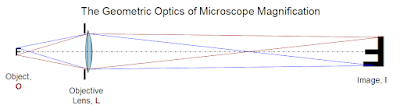 The real image to the right can be seen if you put your eye a suitable distance further to the right, and if you put camera film or a digital sensor in the real image's location, the "prime focal plane" it will record the image. In the arrangement shown, the real image is four times the size of the object.
To analyze the detail we might be able to record, there are two regions of interest:
How much detail can the human eye see? People who are not nearsighted typically look at something no closer than 10" (250mm). Normal vision has an angular resolution (at the point of attention) of about a minute of arc, which means we can resolve items no finer than 1/3,500 of the distance from the eye. Vision called 20/20 is based on resolution closer to 1/1,000, or about 3.5 minutes of arc. Photographs printed at your local drug store have a resolution of 200 pixels per inch, or 8/mm. Held at a close distance, where they still "look pretty good", the finest details are about 1/2,000 the distance to the eye. Let us use this ratio to understand what we need to do to see everything the objective lens above is putting into the real image it presents to us.
If you place your eye ten inches from the real image, you can see it. The smallest details visible to you will be about 1/8mm or 1/200 inch apart; 1/8mm is 125µ. The original 4x system is producing a real image with details as small as about 6µ, so a lot of the detail in the image is wasted.
That is why the compound microscope has an eyepiece. A 10x eyepiece is placed one inch (25mm) from the real image and lets you see the image ten times larger. In this instance, you can now see details in the image as small as 12.5µ. To see "everything that is there", you need a 20x eyepiece, which lets you see 6.25µ details, which is getting close. At that point, the effective magnification is 80x. So how do microscopes with effective magnification of 1000x or so work?
First, we have to get a lens that has a lot smaller Objective focal ratio, smaller than f/1. It was discovered about a century ago that there is a point inside a glass sphere for which spherical aberration was nullified. Simply speaking, that means an easy-to-produce lens can magnify a lot. To get the object "inside" a spherical lens, one side of the sphere is ground flat, and an immersion medium such as oil is used to "extend" the lens so it surrounds the object:
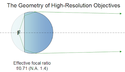 Ratios as low as f/0.55 are possible, but f/0.71 is most common with glasses that need minimal color correction. At f/0.71, the Rayleigh criterion at 0.6µ means that detail on the Object can be seen as small as 1.22x0.6µx0.71 = 0.52µ. What magnification is then possible? Recall that the 1/2,000 criterion means that human eyes can see 1/8mm or 125µ. 125/0.52 = 240x. That is the maximum "useful" magnification using visible light.
Most high-power oil-immersion lenses are designed to provide a real image 100x larger than the area of the object being examined. So when you use a 10x eyepiece, you're seeing that area magnified 1,000x. However, if you examine the image critically (I have done so), you will realize that the edges between light and dark objects do not appear knife-sharp. Try a lower-power eyepiece: 5x looks a lot sharper, though of course everything appears smaller. You can't get 2.5x eyepieces, but if you could, the image you see would be well matched to the ability of your eye. The value of looking at the larger, blurrier image is that you can scan it more effectively for the subtle details, which might be missed at a lower magnification. As a reference, an E. coli cell has a diameter near 2µ and a length of about 5µ, so these critters look pretty small even at 1,000x.
A bit of extra resolution can be had by using a shorter wavelength. I've used, with little explanation, an effective wavelength of 0.6µ. That is the effective wavelength for incandescent light, which is rather orange, though we get used to feeling that it is "white enough". But if we throw away most (not all) of the red-orange light with a filter, we'll have a bluish light that is also "white enough" but has a shorter effective wavelength. My microscope came with a "daylight" filter that shifts the effective wavelength to about 0.48µ. With this filter, the ultimate resolution of the 100x objective is not 0.52µ but 0.42µ, allowing a magnification at maximum sharpness of 300x, and quite a bit more detail is then visible at 1,000x.
A slightly mathematical note to finish this portion: Microscopes have been standardized for decades at a 160mm "tube length", which fixes the distance between the real image and the objective lens at 160mm. The actual size of the microscope depends on the way the lenses are attached. A f/0.71 objective lens (N.A. 1.40) thus has a distance from the object (the microscope slide) of 1.6mm and a diameter of 2.25mm. The lens-to-image focal ratio is 160/2.25 or f/71 (100 times f/0.71). With my blue filter in place, the pixel size of the real image is 42µ (100 times 0.42µ). If I put a digital camera sensor in the focal plane, where the real image is, a lot of its resolution will be wasted. That is why microscopes set up for photography use a relay lens that shrinks the image, usually by a factor of 8, so that it is well-suited to recording by a sensor whose pixels are in the 5µ-10µ range.
These are the limits to seeing fine detail with visible light. Though I'd intended to explore shorter wavelength light (and other radiations), this post is long enough already.
In the prior post I explained that larger eyes can see, and larger cameras can record, more detail from a particular object or scene. Briefly, the size of the smallest spot that can be produced by a diffaction-limited lens has a radius expressed by the Airy formula,
x = 1.22 λf/d
Given an effective wavelength (λ) for "average" visible light of 0.6µ, this formula tells us that the radius x of the smallest spot (on film or digital sensor) that the lens can make depends only on its focal ratio f/d. A small lens with a short focal length can cast an image on a only a small area, comparable to the lens-to-sensor distance. A larger lens at a greater distance from the sensor can cast a useful image on a sensor of larger area. Since the spot size, which limits the useful pixel resolution, is the same for both lenses, the larger lens will capture more detail from the object being viewed or imaged.
Continuing with visible light, let us consider how to see finer and finer detail in extreme close-up mode. When the lens gets closer to the object than it is to the sensor, things get a bit more complex, but only a bit. In the diagram below, the small "F" on the left is being magnified by a lens to produce a real image, shown as a larger "F".
 The real image to the right can be seen if you put your eye a suitable distance further to the right, and if you put camera film or a digital sensor in the real image's location, the "prime focal plane" it will record the image. In the arrangement shown, the real image is four times the size of the object.
To analyze the detail we might be able to record, there are two regions of interest:
- The Object-to-Lens distance and Lens diameter establish the Objective focal ratio, which determines the smallest detail on the Object that can be imaged. In microscopy, this is usually expressed as its inverse, the Numerical Aperture, or N.A. For this diagram, the Objective focal ratio is 2, so the N.A. is 0.5. Using the formula above, the smallest detail seen will have a radius of 1.22x0.6µx2 = 1.46µ.
- The Lens-to-Image distance and Lens diameter establish the Image Focal Ratio, which determines the effective pixel resolution of the system. The magnification here is 4, meaning the lens-to-image distance is 8 times its diameter, so this is a f/8 system. The blur circle is thus 1.22x0.6µx8 = 5.86µ, or about 6µ in radius.
How much detail can the human eye see? People who are not nearsighted typically look at something no closer than 10" (250mm). Normal vision has an angular resolution (at the point of attention) of about a minute of arc, which means we can resolve items no finer than 1/3,500 of the distance from the eye. Vision called 20/20 is based on resolution closer to 1/1,000, or about 3.5 minutes of arc. Photographs printed at your local drug store have a resolution of 200 pixels per inch, or 8/mm. Held at a close distance, where they still "look pretty good", the finest details are about 1/2,000 the distance to the eye. Let us use this ratio to understand what we need to do to see everything the objective lens above is putting into the real image it presents to us.
If you place your eye ten inches from the real image, you can see it. The smallest details visible to you will be about 1/8mm or 1/200 inch apart; 1/8mm is 125µ. The original 4x system is producing a real image with details as small as about 6µ, so a lot of the detail in the image is wasted.
That is why the compound microscope has an eyepiece. A 10x eyepiece is placed one inch (25mm) from the real image and lets you see the image ten times larger. In this instance, you can now see details in the image as small as 12.5µ. To see "everything that is there", you need a 20x eyepiece, which lets you see 6.25µ details, which is getting close. At that point, the effective magnification is 80x. So how do microscopes with effective magnification of 1000x or so work?
First, we have to get a lens that has a lot smaller Objective focal ratio, smaller than f/1. It was discovered about a century ago that there is a point inside a glass sphere for which spherical aberration was nullified. Simply speaking, that means an easy-to-produce lens can magnify a lot. To get the object "inside" a spherical lens, one side of the sphere is ground flat, and an immersion medium such as oil is used to "extend" the lens so it surrounds the object:
 Ratios as low as f/0.55 are possible, but f/0.71 is most common with glasses that need minimal color correction. At f/0.71, the Rayleigh criterion at 0.6µ means that detail on the Object can be seen as small as 1.22x0.6µx0.71 = 0.52µ. What magnification is then possible? Recall that the 1/2,000 criterion means that human eyes can see 1/8mm or 125µ. 125/0.52 = 240x. That is the maximum "useful" magnification using visible light.
Most high-power oil-immersion lenses are designed to provide a real image 100x larger than the area of the object being examined. So when you use a 10x eyepiece, you're seeing that area magnified 1,000x. However, if you examine the image critically (I have done so), you will realize that the edges between light and dark objects do not appear knife-sharp. Try a lower-power eyepiece: 5x looks a lot sharper, though of course everything appears smaller. You can't get 2.5x eyepieces, but if you could, the image you see would be well matched to the ability of your eye. The value of looking at the larger, blurrier image is that you can scan it more effectively for the subtle details, which might be missed at a lower magnification. As a reference, an E. coli cell has a diameter near 2µ and a length of about 5µ, so these critters look pretty small even at 1,000x.
A bit of extra resolution can be had by using a shorter wavelength. I've used, with little explanation, an effective wavelength of 0.6µ. That is the effective wavelength for incandescent light, which is rather orange, though we get used to feeling that it is "white enough". But if we throw away most (not all) of the red-orange light with a filter, we'll have a bluish light that is also "white enough" but has a shorter effective wavelength. My microscope came with a "daylight" filter that shifts the effective wavelength to about 0.48µ. With this filter, the ultimate resolution of the 100x objective is not 0.52µ but 0.42µ, allowing a magnification at maximum sharpness of 300x, and quite a bit more detail is then visible at 1,000x.
A slightly mathematical note to finish this portion: Microscopes have been standardized for decades at a 160mm "tube length", which fixes the distance between the real image and the objective lens at 160mm. The actual size of the microscope depends on the way the lenses are attached. A f/0.71 objective lens (N.A. 1.40) thus has a distance from the object (the microscope slide) of 1.6mm and a diameter of 2.25mm. The lens-to-image focal ratio is 160/2.25 or f/71 (100 times f/0.71). With my blue filter in place, the pixel size of the real image is 42µ (100 times 0.42µ). If I put a digital camera sensor in the focal plane, where the real image is, a lot of its resolution will be wasted. That is why microscopes set up for photography use a relay lens that shrinks the image, usually by a factor of 8, so that it is well-suited to recording by a sensor whose pixels are in the 5µ-10µ range.
These are the limits to seeing fine detail with visible light. Though I'd intended to explore shorter wavelength light (and other radiations), this post is long enough already.
Sunday, May 31, 2009
It takes big eyes to see fine detail
kw: musings, physics, light, visibility
After reviewing The Lightness of Being recently (post here), I began to consider how it takes such large machines to study the tiniest things. Then I spoke with someone who was asking about diffraction, and I explained that it is a visible manifestation of Heisenberg's uncertainty principle.
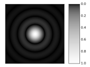 [Airy Disk, image from the Wikipedia Commons. For a discussion, click here.] This shows the blur circle that results when parallel light of a single wavelength passes through a circular opening and is then focused to a "point" by a perfect lens. The radius of the first dark ring is given by the formula

The ratio f/d is just the focal ratio, or f-number of the lens. What this firstly shows is that the absolute size of the Airy Disk is independent of the size of the lens, for a given ration of focal length f to lens diameter d. Thus, when your camera reports it is using f/4 to record a scene, the smallest dot its lens can make on the film or digital sensor is 2.44 times 4 times the brightest wavelength. If we use 0.6μ (600 nanometers, nm) as the "effective" wavelength for whitish light, that smallest dot is about 5.86μ. Let's call it 6μ. If you work out the uncertainty in the position of a photon that results from a given diameter lens, the Heisenberg formula will give the same answer, about 6μ when the ratio of the lens diameter to its distance from the recording medium is 1/4.
After reviewing The Lightness of Being recently (post here), I began to consider how it takes such large machines to study the tiniest things. Then I spoke with someone who was asking about diffraction, and I explained that it is a visible manifestation of Heisenberg's uncertainty principle.
 [Airy Disk, image from the Wikipedia Commons. For a discussion, click here.] This shows the blur circle that results when parallel light of a single wavelength passes through a circular opening and is then focused to a "point" by a perfect lens. The radius of the first dark ring is given by the formula
The ratio f/d is just the focal ratio, or f-number of the lens. What this firstly shows is that the absolute size of the Airy Disk is independent of the size of the lens, for a given ration of focal length f to lens diameter d. Thus, when your camera reports it is using f/4 to record a scene, the smallest dot its lens can make on the film or digital sensor is 2.44 times 4 times the brightest wavelength. If we use 0.6μ (600 nanometers, nm) as the "effective" wavelength for whitish light, that smallest dot is about 5.86μ. Let's call it 6μ. If you work out the uncertainty in the position of a photon that results from a given diameter lens, the Heisenberg formula will give the same answer, about 6μ when the ratio of the lens diameter to its distance from the recording medium is 1/4.
Most point-and-shoot digital cameras these days have pixels of 3μ x 3μ or smaller, which is why these small cameras try to use larger lens openings in the range f/2.5-f/3. The sensors in my digital SLR are closer to 7μ x 7μ, because the sensor is larger. That allows me to use smaller lens openings for some depth of field and still have a lot of detail in the image. This camera is more than twice the size and ten times the weight of a more ordinary pocket camera.
OK, I am getting ahead of myself here. I wrote above that the dot size depends only on the f/number of the lens. That is just the beginning of the story. Let's consider an f/4 system, because we have worked out the relevant figures just above. The blur circle is about 6μ in diameter. The "Rayleigh criterion" for resolving detail states that the center of one spot must be no closer to the center of the next than the radius of the blur circle. In such a situation, one bright center is on top of the dark ring of the next center. Two such spots in an f/4 system are thus 3μ apart.
Now let us consider three possible "eyes" with a focal ratio of f/4. First, we'll discuss a camera with a lens-and-sensor arrangement similar to the human eye in size. This is the situation with many pocket digital cameras. The sensor might record 10 mpx (megapixels), or 3650x2740, and have a size of 6mm x 4.6mm (6,000μ x 4600μ). The individual pixels are only 1.6μ x 1.6μ. The lens focal length for "normal" focus is about the size of the image sensor's diagonal dimension, or 8mm (8,000μ). At f/4, the lens diameter is 2mm (2,000μ) (The lens is bigger than this, allowing it to record images whose blur circle is closer to the size of the image pixels, but it can "stop down" to f/6 or f/8 to exclude excess light or allow greater depth of field).
No matter what the size of the pixels is, the smallest details that can be resolved are in the range of 3μ apart on the sensor. Effectively, 6,000/3 = 2,000 and 4,600/3 = 1,533, such that the true image "detail size" is 3mpx. Only for focal ratios near f/2 can you capture all the detail that sensor can resolve.
Now let us consider a "spider eye", with a lens-to-retina distance of about a millimeter. I picked a spider's eye because it is not segmented like the eyes of most insects, but is a tiny lens-and-retina eye. If such an eye has a lens diameter of 1/4mm (250μ), it will also be able to resolve details that are 3μ apart on the retina. But the largest possible retina that can be kept a millimeter from the lens has a size of less than 3mm x 3mm. Let us suppose the "sweet spot" or middle region is 1.5mm x 1.5mm (1500μ x 1500μ). Let us further assume that the nerve cells in the retina are smaller than 3μ (they are probably close to 2μ in diameter). Then it will be, in effect, a 500x500 pixel array, or about a quarter megapixel. That is sufficiently detailed for many uses, but is actually a bit blurrier than analog TV. Actually, the eye of a real spider uses a spherical lens almost in contact with its retina, so its focal ratio is f/1 or even smaller. This allows for more detailed seeing than an f/4 eye at such a small size, and works in darker environments.
Now let us consider a large landscape camera, of the type often called Graphic; Graflex was a favorite older brand of large camera. These typically were a foot long or more, and recorded on film sizes of 4"x5" (100mm x 125mm) or larger. Such cameras are still made, and some photographers still use large sheet film in them. They make stunning images. One can also get scanning backs for them to record digital landscapes, but they are very costly. How will such a costly system perform at f/4?
Parameters: Focal length about 12" or 300mm, diameter 3" or 75mm. A scanning back or film cassette size of 125mm x 100mm (125,000μ x 100,000μ) is commonly used with a lens of this length. Scanning backs have pixel sizes ranging from 5μ to 10μ, because they have resolution aplenty. A 100mm scanner with a 125mm travel and 10μ pixels will be using each pixel to the fullest. Its final image size is 12,500x10,000, or 125Mpx. That is more than forty times the detail than what we found for the digital camera considered above, and hundreds of times the detail that can be captured by the spider eye.
Now suppose you are using these three systems to look at an object just one meter from the camera lens, and about half a meter (500mm) across. The spider eye will cover the item with 500 pixels (or fewer) each way, seeing details no smaller than a millimeter. The small digital camera will record an image that is effectively 2000 pixels the large way, seeing details as small as 1/4 millimeter. This is about how well your eye does from half a meter away also. The Graphic camera will record near-microscopic details, with 12,500 x 10,000 pixels, recording details smaller than 1/20 mm, or 50μ.
Just considering visible light, it becomes clear that larger cameras are needed to record finer details. To see smaller details than this, or to resolve smaller features of far-away objects, the same principle holds, as I'll get into in the next day or two.
Subscribe to:
Posts (Atom)

 Posts
Posts

